Ureter Trauma

Ureter Trauma

David Ray Velez, MD

Table of Contents

Background

Ureter Injuries in Trauma are Rare

Most Common (95%) After Penetrating Abdominal Trauma

Ureter Trauma is One of the Most Common Sites of Missed Injury at Laparotomy (Rate: 11%)

Missed Injury is Associated with Significant Morbidity (Renal Failure and Sepsis) and Mortality

AAST Ureter Injury Scale

Diagnosis

Presentation

  • Hematuria – An Important Sign but Unreliable (Absent in 67% of Blunt Abdominal Trauma and 45% of Penetrating Abdominal Trauma)
  • Flank Pain
  • Delayed Signs:
    • Persistent Pain
    • Fever
    • Sepsis
    • Fistula
    • Ileus

Presentation is Often Nonspecific and Diagnosis Requires a High Index of Suspicion

Diagnosis

  • May Be Diagnosed Intraoperatively or on Radiographic Imaging
  • Radiographic Imaging:
    • CT with IV Contrast (Need Delayed Excretory Phase) is Most Common
    • May Also Consider an IV Pyelogram (IVP)
    • Consider Cystoscopy with Retrograde Pyelography if Needed
  • Intraoperative Evaluation:
    • Most Often Seen by Direct Visualization
    • IV Methylene Blue or Indigo Carmine May Assist

Proximal Ureter Injury with Extravasation (Left); Delayed Imaging (Right) 1

Treatment

The Primary Treatment for Ureter Injury is Surgical Repair

Timing of Repair

  • Early Diagnosis (< 3-5 Days): Immediate Repair
  • Delayed Diagnosis (> 3-5 Days): Immediate vs Delayed Repair (Controversial)
    • Temporize with Endoscopic Stenting vs Percutaneous Nephrostomy Tube if Indicated When Proceeding with Delayed Repair
    • Developing Inflammation, Edema, and Friability Increase the Risk of Complications if Attempting Immediate Repair After a Delayed Diagnosis

Definitive Repair

  • Upper 2/3 (Over Pelvic Brim): Ureteroureterostomy (Primary Anastomosis)
  • Lower 1/3 (Below Pelvic Brim): Ureteroneocystostomy (Reimplant in Bladder)
  • *See Techniques Below

Damage Control Options

  • Cutaneous Ureterostomy – Stent Placed Through the Proximal Transected Ureter and Externalized to Control Urinary Output Until Stabilized for Delayed Definitive Repair
  • Ligate Ends with a Percutaneous Nephrostomy

Surgical Technique

Always Use Absorbable Sutures – Avoid Stricture/Stones

Avoid Skeletonization When Dissecting – Risk for Devascularization
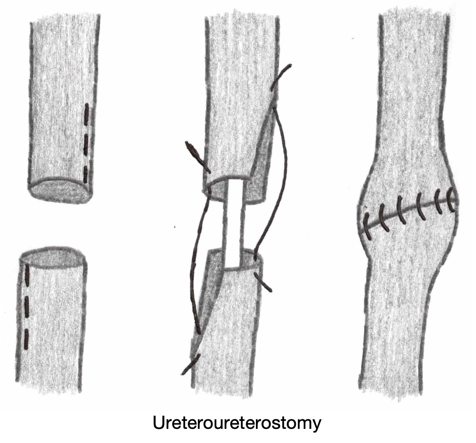
Ureteroureterostomy

  • Definition: Ureter-to-Ureter Anastomosis
  • Debride Devitalized Tissue
  • Spatulate the Proximal and Distal Ends
  • Insert a Double-J Ureteral Stent
  • Create the Anastomosis Over the Stent
    • Use Absorbable Monofilament Sutures (PDS 5-0) to Avoid Stricture/Stones
  • *Large Injuries May Require Kidney Mobilization and Nephropexy to Decrease Tension

Ureteroneocystostomy

  • Definition: Ureter Reimplantation into the Bladder
  • Debride Devitalized Tissue
  • Spatulate the Proximal End of the Ureter
  • Create a Small Cystotomy
  • Insert a Double-J Ureteral Stent
  • Anastomose the Ureter to the Bladder Cystotomy Over the Stent
    • Use Absorbable Monofilament Sutures (PDS 5-0) to Avoid Stricture/Stones

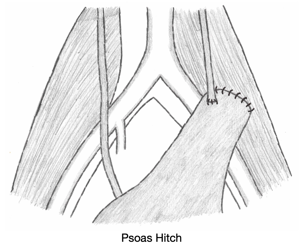
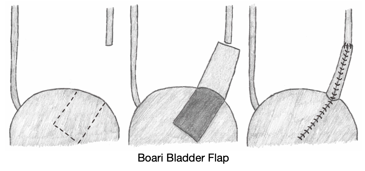
Adjuncts to Decrease Tension for Large Defects

  • Psoas Hitch– Mobilize Bladder and Anchor to the Psoas (Generally Preferred)
  • Bladder (Boari) Flap – Bladder is Tubularized
  • Transureteroureterostomy (TUU) – Implant into the Contralateral Ureter
  • Ileal Interposition
  • Renal Autotransplantation – Autotransplant of the Ipsilateral Kidney into the Pelvis

References

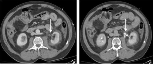
  1. Ortega SJ, Netto FS, Hamilton P, Chu P, Tien HC. CT scanning for diagnosing blunt ureteral and ureteropelvic junction injuries. BMC Urol. 2008 Feb 7;8:3. (License: CC BY-2.0)