Vascular Exposure: Subclavian

Vascular Exposure: Subclavian

David Ray Velez, MD

Table of Contents

Anatomy/Branches

First Part: Proximal to the Anterior Scalene

  • Vertebral Artery
  • Internal Thoracic Artery (Internal Mammary Artery)
    • Anterior Intercostal Arteries
    • Becomes the Superior Epigastric Artery
  • Thyrocervical Trunk
    • Inferior Thyroid Artery
    • Suprascapular Artery
    • Transverse Cervical Artery

Second Part: Behind the Anterior Scalene

  • Costocervical Trunk
    • Superior Intercostal Artery
    • Deep Cervical Artery

Third Part: Distal to Anterior Scalene

  • Dorsal Scapular Artery

Termination: Becomes the Axillary Artery Past the First Rib

Right Subclavian Vessels

Proximal Access: Median Sternotomy

Distal Access: Supraclavicular Incision (*See Technique Below)

May Consider a “Hockey Stick” Incision with a Median Sternotomy and Supraclavicular Extension if Access to the Entire Right Subclavian Vessel is Required

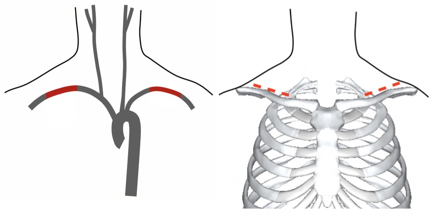
Exposure by Median Sternotomy

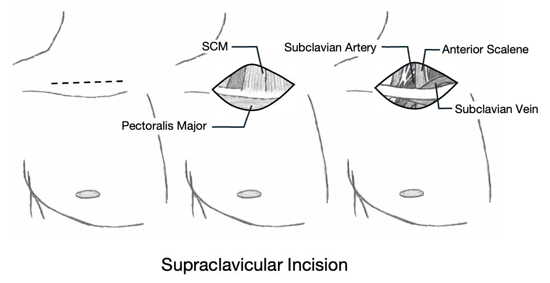
Exposure by Supraclavicular Incision

Left Subclavian Vessels

The Left Subclavian Vessels are Generally Considered the Most Difficult to Expose

Proximal Access Options:

  • Anterior Left Thoracotomy Through the Third Intercostal Space
    • Exposure is Often Limited
  • Resuscitative Anterolateral Left Thoracotomy
  • Median Sternotomy with Supraclavicular Extension
    • Median Sternotomy Alone is Generally Inadequate to Access the Left Subclavian Due to the Deep Position
  • “Trap Door” Incision
    • Three Components:
      • Anterolateral Left Thoracotomy
      • Partial Sternotomy
      • Left Supraclavicular Incision
    • The “Door” Can Then Be Rotated Out to Allow Access to the Deeper Structures
    • Considered the Classical Incision but is Associated with Significant Morbidity, More Severe Bleeding, and Respiratory Complications
    • Generally Not Used in Modern Practice
  • Clavicular Resection
    • May Require Addition of Median Sternotomy to Improve Proximal Access

Distal Access: Supraclavicular Incision (*See Technique Below)

  • Artery is Deeper and More Difficult to Expose than on the Right-Side
  • Can Resect the Clavicle to Improve Access

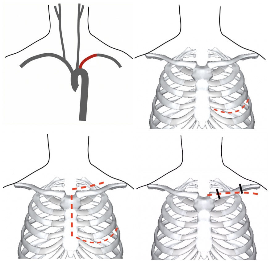
Access to the Left Subclavian (Thoracotomy, Trap Door, and Clavicular Incision)

Supraclavicular Incision

Allows Access to the Mid-Distal Subclavian Artery and Vertebral Artery (Segment V1)

Incision

  • Traditional Incision: Transverse Incision 1 cm Above the Clavicle
  • If Planning to Preform Clavicular Resection from the Start: Transverse Incision Over the Anterior Surface of the Clavicle from the Sternoclavicular Junction to the Mid-Clavicle and Down Over the Deltopectoral Groove

Technique

  • Carry Incision Down Through the Platysma
  • Divide the Clavicular Attachment of the SCM
    • Exposes the Internal Jugular Vein and Scalene Fat Pad
  • Divide the Anterior Scalene Muscle
    • Exposes the Subclavian Artery
    • The Anterior Scalene Muscle Lies Between the Subclavian Vein (Anterior) and Artery (Posterior)
    • Preserve the Phrenic Nerve Coursing Obliquely Over the Anterior Scalene Muscle
  • Vertebral Artery (Segment V1) Can Be Identified Around the Medial Portion of the Subclavian Artery

Options to Improve Visualization

  • Clavicular Resection
    • Clear Circumferentially of All Tissues
    • Use Gigli Saw to Divide in the Mid Portion
    • Dissect the Sternal Head Free to Remove the Clavicle
    • Can Replace Clavicle When Done (But Not Mandatory)
  • Median Sternotomy
    • Improves Proximal Access
  • Extend Incision Down Over the Deltopectoral Groove
    • Improves Distal Access Toward the Axillary Vessels