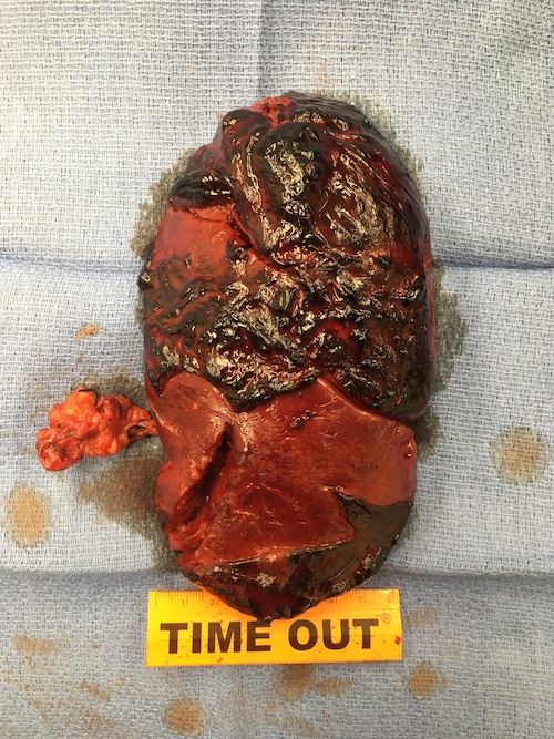
Spleen Trauma
Spleen Trauma
David Ray Velez, MD
Table of Contents
Background
Vasculature
- Splenic Artery: From the Celiac Trunk
- Runs Superior to the Pancreas
- Very Tortuous and Divides into 5-6 Branches
- Splenic Vein: Drains into the Portal System
- Runs Posterior/Inferior to the Artery (Posterior or Within the Pancreas)
Ligaments
- Gastrosplenic Ligament: Hilum to Stomach
- Contains the Short Gastric Vessels
- Splenorenal Ligament (Lienorenal Ligament): Hilum to Left Kidney
- Contains the Splenic Artery/Vein and Tail of the Pancreas
- Splenophrenic Ligament: Superior Pole to Diaphragm
- Avascular
- Splenocolic Ligament: Inferior Pole to Colon
- Avascular
- Phrenicocolic Ligament (Hensing’s Ligament): Diaphragm to Splenic Flexure of the Colon
- Supports Spleen but Not Directly Associated
Physiology/Function
- Red Pulp (75-85%)
- Filters Old/Senescent or Damaged Red Blood Cells
- Traps Bacteria
- White Pulp (15-25%)
- Produces Lymphocytes and Antibodies
Etiology
- Blunt Abdominal Trauma:
- Most Common Organ Injury in Blunt Abdominal Trauma (40-55%)
- *Remains Controversial and Reports Vary Between Spleen and Liver
- Most Common Injury Requiring Intervention in Blunt Abdominal Trauma
- Most Common Organ Injury in Blunt Abdominal Trauma (40-55%)
- Penetrating Abdominal Trauma:
- Less Common After Penetrating Abdominal Trauma
AAST Spleen Injury Scale (2018 Revision)
- *See AAST
- Injury Scale is Under Copyright

Spleen Injury
Initial Evaluation and Management
Unstable: Laparotomy
- First Perform a FAST (Focused Assessment with Sonography in Trauma) to Confirm Abdominal Source
- Diagnostic Peritoneal Lavage (DPL) is Rarely Performed but May Be Considered if FAST is Inconclusive
- Search for Oher Sources if Negative
- Diffuse Peritonitis Indicates Bowel Injury and Warrants Laparotomy – Diffuse Peritonitis Should Never be Attributed to Solid Organ Injury as Isolated Hemoperitoneum Should Not Cause Diffuse Peritoneal Irritation
Stable: CT Imaging
Indications for Angiography and Embolization (93% Success Rate)
- Transient Responder
- Active Extravasation (“Blush”)
- Pseudoaneurysm
- Arteriovenous Fistula (AVF)
Stable Patients Generally Undergo a Trial of Nonoperative Management (With or Without Angioembolization) Unless There are Other Indications for Surgery
Surgery Solely for Washout of Blood is Generally Not Indicated Unless for Severe Refractory Pain
*Historically Severe Traumatic Brain Injury (TBI) and Altered Mental Status (AMS) Were Contraindications to Nonoperative Management of the Spleen (Concern for Hypotension and Secondary Brain Injury) – Now Shown to Be Effective and Safe
Nonoperative Management (NOM)
The Exact Definitions for Nonoperative Management are Poorly Defined
Admission
- Consider ICU Admission for 24-72 Hours for Injuries ≥ Grade III
- Consider an Initial NPO Status for up to 24 Hours if Closely Monitoring
- Exact Hospital Length of Stay is Poorly Defined
Activity Restrictions
- Bedrest: Consider for 24-48 Hours
- Previously Defined as Injury Grade + One Day (1999 APSA Guidelines)
- Activity Restriction: Return to Unrestricted Activity at Injury Grade + 2 Weeks
- Defined as “Normal” Age-Appropriate Activities
- Longer Restrictions for Contact Sports (Football, Wrestling, Hockey, etc.) – Less Well Defined but Often Range from 2-6 Months
Indications for Surgery (After a Trial of Nonoperative Management)
- Angioembolization Failure
- Hemodynamic Instability
- Persistent Bleeding
- Refractory Abdominal Pain
- *May Consider Angiography and Embolization if Stable and Not Already Attempted
Start DVT Prophylaxis Early (Within 24-48 Hours) for Solid Organ Injury if Otherwise Clinically Appropriate
Repeat CTA – Controversial
- Routine Use without Clinical Indication is Not Useful
- Most Studies Show that Routine Use is Not Necessary and Rarely Changes Clinical Management
- Some Advocated for Repeat CTA After 2-7 Days, Before Discharge, to Evaluate for Pseudoaneurysm or Arteriovenous Malformation
Surgical Management
Small and Minimal Bleeding (Grade I): Topical Hemostasis
- Options:
- Manual Compression
- Electrocautery
- Argon Beam Coagulation
- Arista
- Surgical (Original, Powder, Fibrillar, Nu-Knit)
- Fibrin Glue
- Do Not Need to Explore Subcapsular Hematomas if Stable and Not Expanding
Simple Laceration (Grade II-III): Splenorrhaphy (Primary Repair)
- Generally Use Monofilament or Chromic Suture
- Spleen Does Not Hold Suture Well – Consider Pledgets (Felt, PTFE) or Absorbable Mesh
- May Consider Splenectomy vs. Partial Splenectomy Depending on the Severity of Injury and Patient Stability
Shattered or Vascular/Hilar Injury (Grade IV-V): Splenectomy
Vaccination
Loss of Antibody-Mediated Immunity to Capsulated Bacteria in the Spleen Creates a Risk for Overwhelming Post-Splenectomy Infection (OPSI)
Vaccinations
- Hemophilus influenzae Type B (HiB) – Single Dose Only
- Meningococcus (MCV) – Multiple Doses with a Booster After 5 Years
- Pneumococcus (PCV) – Single Dose with a Booster After 5 Years
Timing
- Elective Splenectomy: Finish Course 2 Weeks Before (Start 10-12 Weeks Prior)
- Emergent Splenectomy: Start Course 2 Weeks After (Impaired Functional Antibody Responses Prior)
- Give Just Prior to Discharge if Unreliable or Concerned for Loss to Follow Up
Always Recommended After Splenectomy
Recommend Against Routine Vaccination After Splenic Artery Embolization in Adults – Fewer Infectious Complications and Greater Preservation of Splenic Immune Function
