POCUS: Left Ventricular Ejection Fraction (LVEF)
POCUS: Left Ventricular Ejection Fraction (LVEF)
David Ray Velez, MD
Table of Contents
Left Ventricular Ejection Fraction (LVEF)
Definition
- LVEF = SV/EDV x 100
- SV: Stroke Volume
- EDV: End-Diastolic Volume
- Ejection Fraction is a Measure Used in Evaluating Heart Function
Severity
- > 70%: Hyperdynamic
- 50-70%: Normal
- 40-49%: Mildly Reduced
- 30-39%: Moderately Reduced
- < 30%: Severely Reduced
Qualitative Assessment
- Left Ventricle Wall Movement
- Minimal Movement Indicates Reduced EF
- Near-Collapse Indicates Hyperdynamic EF
- Anterior Mitral Valve Movement
- Should Move Toward the Ventricular Septum and Nearly Touch
- Minimal Movement Toward the Ventricular Septum Indicates Reduced EF
Quantitative Assessment
- E-Point Septal Separation (EPSS): Estimates LVEF By Assessing Mitral Valve Leaflet Movement Toward the Ventricular Septum
- Fractional Shortening: Estimates LVEF By Assessing Change in Diameter of the LV
- Fractional Area Change: Estimates LVEF By Assessing Change in Cross-Sectional Area of the LV
- Simpson (Biplane) Method: Estimates LVEF By Assessing Change in Volume of the LV
POCUS Cardiac Views
- Using a Phased-Array Probe
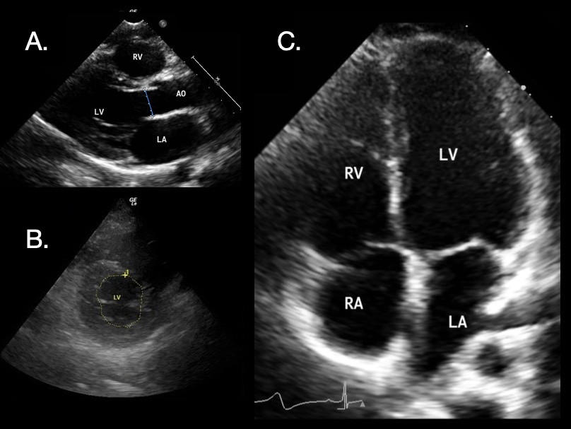
- Parasternal Long-Axis View (PLAX): Left Sternal Border at the 3rd-4th Intercostal Space, Probe Indicator to the Right Shoulder
- Parasternal Short-Axis View (PSAX): Left Sternal Border at the 3rd-4th Intercostal Space, Probe Indicator to the Left Shoulder
- Apical 4-Chamber View (A4C): Just Inferior to the Left Nipple, Probe Indicator to the Left Flank
- Subcostal View: 2-3 cm Below the Xyphoid
- *See Point-of-Care Ultrasound (POCUS)

Cardiac POCUS: (A) Parasternal Long-Axis 1; (B) Parasternal Short-Axis 2; (C) Apical 4-Chamber 1 [Right Atrium (RA), Right Ventricle (RV), Left Atrium (LA), Left Ventricle (LV), Aortic Outflow (AO)]
E-Point Septal Separation (EPSS)
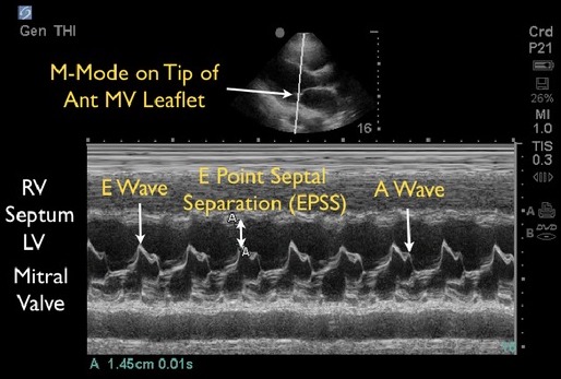
E-Point Septal Separation (EPSS): Estimates LVEF By Assessing Mitral Valve Leaflet Movement Toward the Ventricular Septum
Technique
- Parasternal Long-Axis View (PLAX)
- M Mode Over the Distal Anterior Leaflet of the Mitral Valve
- EPSS is the Closest Distance of the Mitral Valve to the Septum (The Largest Peak Seen in M Mode)
Interpretation
- EPSS > 7 mm Indicates Reduced LVEF
- EPSS > 13-18 mm Indicates Severely Reduced LVEF
- LVEF = 75.5 – (2.5 x EPSS)
- *Not Valid with Mitral Valve (Stenosis, Regurgitation, Repair)

EPSS Measured by POCUS: E Wave (Early Filling of the LV by Passive Blood Flow from the LA), A Wave (Atrial Kick) 3
Fractional Shortening
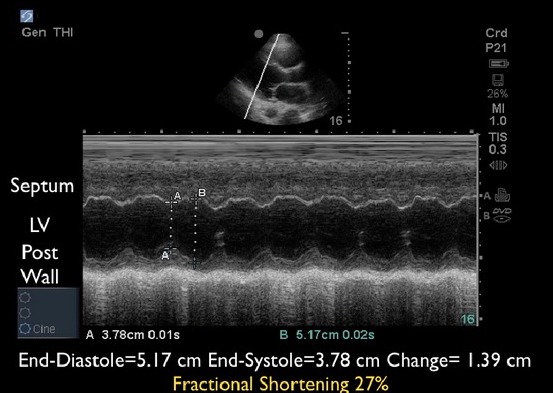
Fractional Shortening: Estimates LVEF By Assessing Change in Diameter of the LV
Technique
- Parasternal Long-Axis View (PLAX)
- M Mode in the Middle of the Left Ventricle
- Left Ventricular End-Diastolic Diameter (LVEDD) – Largest Diameter
- Used to Approximate Diastolic Volume
- Vd = [7/(2.4 + LVEDD)] x LVEDD3
- Left Ventricular End-Systolic Diameter (LVESD) – Smallest Diameter
- Used to Approximate Systolic Volume
- Vs = [7/(2.4 + LVESD)] x LVESD3
Interpretation
- LVEF = (Vd – Vs)/Vd
- Many Ultrasound Machines Will Automatically Calculate
- *May Be Limited by Arrhythmias or Wall Motion Abnormalities

Fractional Shortening on POCUS 3
Fractional Area Change (FAC)
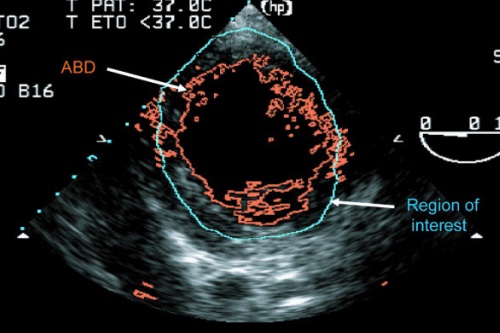
Fractional Area Change: Estimates LVEF By Assessing Change in Cross-Sectional Area of the LV
Technique
- Parasternal Short-Axis View (PSAX)
- LV Area Noted While Viewing Mitral Valve Function at the Midpapillary Muscle
- Left Ventricular End-Diastolic Area (LVEDA) – Largest Area
- Left Ventricular End-Systolic Area (LVESA) – Smallest Area
Interpretation
- FAC (%) = (LVEDA-LVESA)/LVEDA x 100
- 60% FAC: 75% LVEF
- 50% FAC: 66% LVEF
- 40% FAC: 54% LVEF
- 30% FAC: 42% LVEF
- 20% FAC: 29% LVEF
- 10% FAC: 15% LVEF
- *May Be Limited by Wall Motion Abnormalities

Fractional Area Change (FAC) on POCUS 4
Simpson (Biplane) Method
Simpson (Biplane) Method: Estimates LVEF By Assessing Change in Volume of the LV
Technique
- Requires Multiple Views to Estimate LV Volume in 3-Dimensions
- Left Ventricular End-Diastolic Volume (LVEDV) – Largest Diameter
- Left Ventricular End-Systolic Volume (LVESV) – Smallest Diameter
Interpretation
- LVEF = (LVEDV-LVESV)/LVEDV x 100
- *The Most Accurate but the Most Technically Challenging
References
- Gaspar HA, Morhy SS. The Role of Focused Echocardiography in Pediatric Intensive Care: A Critical Appraisal. Biomed Res Int. 2015;2015:596451. (License: CC BY-3.0)
- Mok KL. Make it SIMPLE: enhanced shock management by focused cardiac ultrasound. J Intensive Care. 2016 Aug 15;4:51. (License: CC BY-4.0)
- Seif D, Perera P, Mailhot T, Riley D, Mandavia D. Bedside ultrasound in resuscitation and the rapid ultrasound in shock protocol. Crit Care Res Pract. 2012;2012:503254. (License: CC BY-3.0)
- Cannesson M, Slieker J, Desebbe O, Farhat F, Bastien O, Lehot JJ. Prediction of fluid responsiveness using respiratory variations in left ventricular stroke area by transoesophageal echocardiographic automated border detection in mechanically ventilated patients. Crit Care. 2006;10(6):R171. (License: CC BY-2.0)
Cover Photo: Cannesson M, Slieker J, Desebbe O, Farhat F, Bastien O, Lehot JJ. Prediction of fluid responsiveness using respiratory variations in left ventricular stroke area by transoesophageal echocardiographic automated border detection in mechanically ventilated patients. Crit Care. 2006;10(6):R171. (License: CC BY-2.0)
