Pericardial Effusion

Pericardial Effusion

David Ray Velez, MD

Table of Contents

Introduction

Definition: A Collection of Fluid within the Pericardium Beyond Small Physiologic Amounts (15-50 mL)

Causes

  • Idiopathic
  • Malignancy
  • Uremia
  • Iatrogenic
  • Traumatic
  • Post Myocardial Infarction
  • Acute Pericarditis

Presentation

  • Most are Asymptomatic if Not Hemodynamically Significant
  • Cardiac Tamponade and Hemodynamic Instability
  • Chest Pain
  • Ewart Sign: Dullness (Woody) to Percussion, Egophony, and Bronchial Breath Sounds at the Inferior Angle of the Left Scapula
    • Theoretically Due to Effusion Size Being Large Enough to Compress the Left Lower Lobe of the Lung
    • *More of a Historical Interest Due to Low Sensitivity or Specificity

Size

  • Rapid Accumulation of Even a Small Amount of Blood is Higher Risk for Collapse – Even Larger Volumes Can Be Well Tolerated if They Accumulate Slowly Over Weeks-Years
 VolumeThickness on Echocardiogram
Small< 100 cc< 10 mm
Moderate100-500 cc10-20 mm
Large> 500 cc> 20 mm

Diagnosis

Diagnosis

  • Primarily Diagnosed by Echocardiogram
  • CT or MRI May Be Useful if Echocardiogram is Non-Diagnostic

EKG Findings

  • Sinus Tachycardia
  • Low QRS Voltage
  • Electrical Alternans – Cyclic Beat-to-Beat Shift in the QRS Axis from Mechanical Swinging of the Heart within a Large Effusion

Echocardiogram Findings

Differentiate from Pathology with a Similar Appearance

  • Pericardial Fat Pad – Usually Located Anteriorly and Will Not Be Completely Anechoic (Pericardial Effusion May Be More Circumferential and More Hypoechoic)
  • Pleural Effusion – Will Be Seen Posterior to the Descending Aorta (Pericardial Effusion is Seen Anterior to the Descending Aorta)

Signs of Cardiac Tamponade

  • Systolic Right Atrial Collapse – First Sign
  • Diastolic Right Ventricular Collapse
  • Dilated and Noncollapsible IVC
  • “Swinging Heart” with Pendular Motion from Circumferential Fluid Accumulation

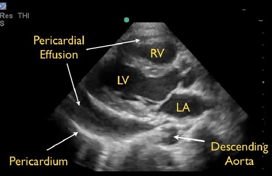
Pericardial Effusion on POCUS: Anterior to the Descending Aorta 2

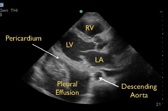
Pleural Effusion on POCUS: Posterior to the Descending Aorta 2

Treatment

Cardiac Tamponade

Pericardial Fluid Drainage

  • Indications:
    • Cardiac Tamponade or Hemodynamic Instability
    • Suspected Purulent Pericarditis (Bacterial or Tuberculosis)
    • Suspected Malignant Pericarditis
    • Large Chronic (> 3 Months) Idiopathic Effusion of Unknown Etiology for Diagnostic Evaluation
    • Symptomatic Moderate-Large Effusion that Fails Medical Therapy
  • Approach:
    • Pericardiocentesis is Generally Preferred
    • Indications for Surgical Median Sternotomy:
      • Concerned for Bleeding/Traumatic Injury
      • Inaccessible to Pericardiocentesis
      • Effusion Small or Loculated
      • Need for Biopsy
      • Coagulopathic

Medical Management

  • Initial Medical Management Alone May Be Appropriate for Even Large Effusions without an Immediate Indication for Drainage
  • Monitor with Serial Exams and Echocardiograms Every 5-7 Days
  • Avoid Volume Depletion
  • Treat Any Underlying Cause

References

  1. COVER: BruceBlaus. Wikimedia Commons. (License: CC BY-3.0)
  2. Seif D, Perera P, Mailhot T, Riley D, Mandavia D. Bedside ultrasound in resuscitation and the rapid ultrasound in shock protocol. Crit Care Res Pract. 2012;2012:503254. (License: CC BY-3.0)