Vascular Exposure: Lower Extremity
David Ray Velez, MD
Table of Contents
Iliac Vessels
Approach
- Common Iliac Artery/Vein (CIA/CIV): Laparotomy
- Internal Iliac Artery/Vein (IIA/IIV): Laparotomy
- External Iliac Artery/Vein (EIA/EIV): Laparotomy (Most Expedient) vs Retroperitoneal Approach
Retroperitoneal Approach
- Curvilinear Incision from the Inguinal Ligament to 2 cm Above the Anterior Superior Iliac Spine (ASIS)
- Carried Down Through the Fat and Subcutaneous Tissue
- Incise the Fascia of the External Oblique and Transversalis
- Pitfalls:
- Avoid Inadvertent Entry into the Peritoneum
- Avoid Injury to the Ureter – Crosses Over the Bifurcation
- Can Extend the Incision Inferiorly (“Hockey Stick”) with Division of the Inguinal Ligament for More Distal Exposure of the Femoral Vessels
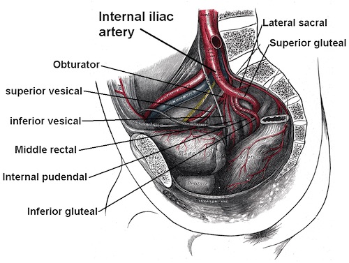
Iliac Vessel Anatomy
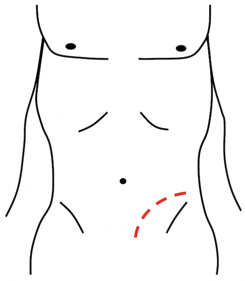
Retroperitoneal Approach for Iliac Vessel Exposure
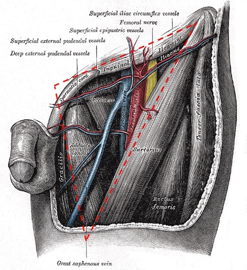
Femoral Vessels
Femoral Triangle
- Borders:
- Inguinal Ligament
- Medial Border of the Sartorius Muscle
- Medial Border of the Adductor Longus Muscle
- Contents (Lateral-to-Medial):
- Femoral Nerve
- Common Femoral Artery (CFA)
- Proximal Superficial Femoral Artery (SFA)
- Proximal Profunda Femoral Artery (PFA)
- Common Femoral Vein
- MNEMONICS for Order of the Femoral Canal Structures
- “NAVEL to the Navel”: Nerve-Artery-Vein-Empty Space-Lymphatics
- “Venous to the Penis”: Vein is Medial to Artery
Femoral Triangle
Proximal Femoral Vessel Exposure
- Incision: Vertical Incision 2-Fingerbredths Lateral to the Pubic Tubercle
- Proximal Extent: Inguinal Ligament
- Approximated by a Line from the Pubic Tubercle to the ASIS
- Caution: Groin Crease Generally Results in the Incision Being Too Low
- Extend Distally Along the Medial Border of the Sartorius Muscle
- Proximal Extent: Inguinal Ligament
- Open the Femoral Sheath to Expose the Common Femoral Vessels and Bifurcation
- Avoid Injury to the Lateral Femoral Circumflex Vein – Crosses Over the Profunda Femoral Artery at the Bifurcation
Can Extend the Proximal Incision Superiorly and Curvilinear to 2 cm Above the ASIS (“Hockey Stick”) with Division of the Inguinal Ligament for Exposure of the External Iliac Vessels
Distal Superficial Femoral Artery (SFA)
- Incision: Along the Medial Border of the Sartorius Muscle
- Retract the Sartorius Medially to Expose Hunter’s Canal
- Unroof Hunter’s Canal to Expose the Distal SFA
- Hunter’s Canal:
- Canal Travelling Under the Sartorius Muscle Containing the Femoral Neurovascular Bundle
- Extends from the Apex of the Femoral Triangle to the Adductor Hiatus
Popliteal Vessels
“Frog-Leg” Position
- Hip Abduction with Slight Lateral Rotation and Knee Flexion
- Bump Under the Knee to Maintain Position
Above-the-Knee Popliteal Exposure
- Incision: Medial Lower Thigh Longitudinally Between the Vastus Medialis and Sartorius Muscles
- The Vascular Sheath is Identified in the Fat Pad Between the Vastus Medialis and Sartorius Muscles
- May Require Division of the Adductor Magnus or Sartorius Muscles
- Popliteal Artery is Encountered First (Medial to the Vein)
Avoid Injury to the Great Saphenous Vein (GSV) with Medial Incisions
Below-the-Knee Popliteal Exposure
- Incision: Longitudinal Medial Incision 1 cm Posterior to the Tibia
- 10 cm Length Starting 1-2 cm Distal to the Medial Femoral Condyle
- Retract the Gastrocnemius Muscle Posteriorly
- Dissect the Soleus Muscle Off the Posterior Tibia
- Popliteal Vein is Encountered First (Medial to the Artery)
- Trifurcation is Identified as the Popliteal Artery Crosses Over the Popliteus Muscle, Between the Gastrocnemius and Soleus
- May Require Division of the Semimembranosus Muscle and Pes Anserinus (Conjoined Tendon of the Sartorius, Gracilis, and Semitendinosus)
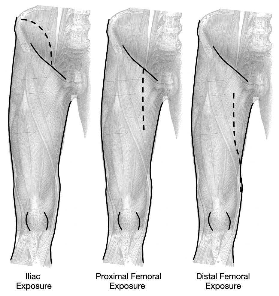
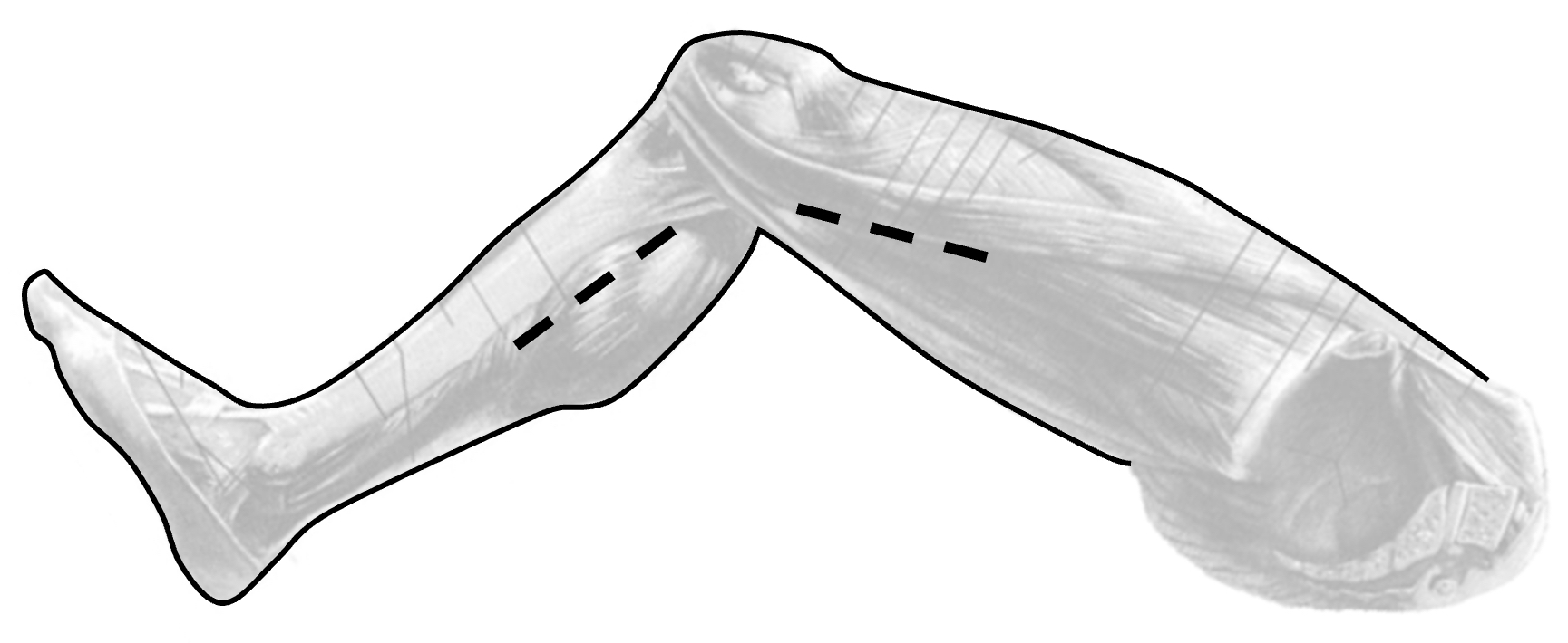
Popliteal Artery Exposure: Above- and Below-the-Knee