Tube Thoracostomy (Chest Tube)
David Ray Velez, MD
Table of Contents
Tube Size
Pneumothorax (PTX): 8-16 French
Pleural Effusion/Hemothorax (HTX): 24-28 French
- *Older Teaching to Use the Largest Size Possible (36-40 French) for Hemothorax Has Fallen Out of Favor – In General Clotted Blood Will Not Drain No Matter How Big the Tube Is
- Use Smaller Sizes in Peds
Excessively Large Sizes Cause Significant Discomfort and Can Inhibit Deep Breathing without Additional Benefit
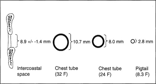
Chest Tube Sizes Compared to Average Intercostal Space (Mid-Axillary 5th Space) 1
Placement Technique
Prepare
- Position Supine with the Ipsilateral Arm Extended or Over the Head
- Prep and Drape Skin
- Inject Local Anesthetic – Inject the Incision Site and the Tract All the Way Down to the Pleura
Incision
- Target 4th/5th Intercostal Space at Anterior Axillary or Mid-Axillary Line
- Make a 2-3 cm Transverse Incision Over the Inferior Rib
- Using Curved Clamp Bluntly Dissect and Tunnel Immediately Above the Rib
- Never Go Below the Rib (Risk Damage to the Neurovascular Bundle)
- Penetrate the Pleura with the Curved Clamp and Spread the Penetration Site
Insertion
- Insert a Finger to Confirm Position and Bluntly Dissect Any Surrounding Adhesions
- Place a Clamp In-Line with the Tip of the Chest Tube
- Insert the Chest Tube Using the Clamp and Direct it into Position
- Pneumothorax (PTX): Aim Anterior/Superior
- Hemothorax (HTX): Aim Posterior
Suture Anchor the Tube to the Skin
Always Obtain a Post-Placement Plan Radiograph to Confirm Placement and Lung Expansion
Alternative Techniques
- Seldinger Technique
- Ultrasound (US)-Guided
- Intervention Radiology (IR)-Guided
Thoracic Irrigation (Thoracic Lavage)
- Definition: Irrigation of the Thorax Through the Chest Tube Upon Insertion
- Used for Hemothorax Only (Not Pneumothorax)
- Reduces the Risk of Retained Hemothorax and Secondary Interventions
- Irrigation Volumes ≥ 1,000 mL Normal Saline are Preferred – Shown Shorter Hospital Length of Stay
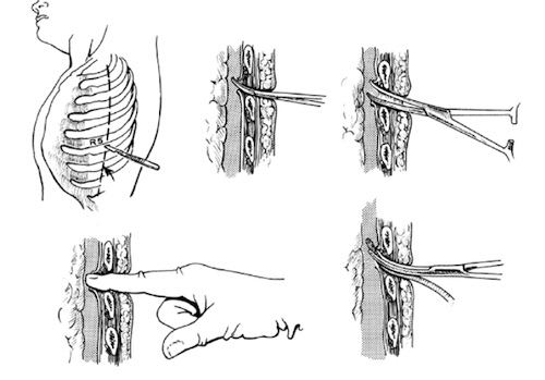
Chest Tube Placement 2
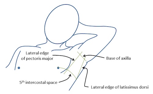
Triangle of Safety 3
Drainage System
Classic “Three Bottle” System
- Bottle #1 (Collecting Bottle): Collects Fluid from the Patient
- Bottle #2 (Water Seal): Allows Air to Escape but Prevents External Air Entry Back into the Chest
- “Bubbling” Indicates an Air Leak
- Bottle #3 (Suction Control): Adjustable Manometer Set to Specific Depths to Set a Controlled Suction Pressure
- Most Often Set to 20 cm H2O
“Four Bottle” System
- Adds an Additional Venting Bottle Connected to the Collecting Bottle
- Fourth Bottle is Vented to the Air and Not Connected to Suction
- Acts as an Additional Water Seal to Prevent Pneumothorax in the Event of Unexpected Suction Failure
Classic “Three Bottle” System 4
Modern Collecting Systems (Atrium, Pleur-Evac)
- Entire System is Combined in a Single Piece of Equipment
- Collecting Chamber Acts as Bottle #1
- Water Seal Acts as Bottle #2
- Suction Control Can be “Wet Suction” or “Dry Suction”
- Wet Suction: Acts as a Traditional Bottle #3 Using Fluid
- Dry Suction: Controls Suction Using a Pressure Control Valve
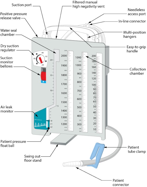
Modern Collecting System 5
Removal
Removal
- Use an Occlusive Dressing to Prevent Inspiration of Air Back into the Pleural Cavity
- Ex: Xeroform or Vaseline-Soaked Gauze
- Cut Any Suture Anchoring to the Skin
- Remove Chest Tube on Expiration, After A Complete Full Inspiration
- Increases Intrathoracic Pressure to Decrease the Risk of Recurrent Pneumothorax Upon Pull
- *Exact Timing is Debated and Some Prefer Pulling with an Inspiratory Hold
Repeat Chest Radiograph
- Consider Repeating Chest Radiograph After Chest Tube Removal (“Post-Pull CXR”)
- Often Done at 4 Hours or 24 Hours
- *No Evidence that Post-Pull CXR is Necessary – Some Prefer Repeat Imaging Based Solely on Symptoms
References
- Gammie JS, Banks MC, Fuhrman CR, Pham SM, Griffith BP, Keenan RJ, Luketich JD. The pigtail catheter for pleural drainage: a less invasive alternative to tube thoracostomy. JSLS. 1999 Jan-Mar;3(1):57-61. (License: CC BY-NC-ND-3.0)
- Mohammed HM. Chest tube care in critically ill patient: A comprehensive review. Egyptian Journal of Chest Diseases and Tuberculosis. 2015. 64(4):849-855. (License: CC BY-NC-ND-4.0)
- Hill J. Taming the SRU. (License: CC BY-NC-SA-3.0)
- Shi H, Mei L, Che G. [The current concepts of closed chest drainage in lobectomy of lung cancer]. Zhongguo Fei Ai Za Zhi. 2010 Nov;13(11):999-1003. (License: CC BY-4.0)
- British Columbia Institute of Technology (BCIT). Wikimedia Commons. (License: CC BY-SA-4.0)