Cardiac Laceration

Cardiac Laceration

David Ray Velez, MD

Table of Contents

Presentation

The Majority of Penetrating Cardiac Injuries are Simple Lacerations

Penetrating Cardiac Trauma Has High Mortality – Only 25-50% Survive to the Hospital

Chambers

  • Right Ventricle is the Most Common Injured Chamber (Due to Anterior Position)
  • Left Atrium is the Least Common Injured Chamber (Due to Posterior Position)

Muscular Ventricle May Seal Lacerations Preventing Exsanguination Prior to Arrival

Most Common Vessel Injured: Left Anterior Descending (LAD)

Coronary Artery Injury Can Cause Cardiac Ischemia and Myocardia Infarction (MI)

Cardiac Tamponade

  • Stab Wounds – Up to 80 will Eventually Cause Tamponade
  • Gun Shot Wounds – Have Larger Pericardial Defects and are More Commonly Associated with Hemorrhage than Tamponade
  • Freely Bleeding Coronary Arteries Can Cause Tamponade
  • Development of Pericardial Tamponade May Have a Protective Effect from Exsanguination and Has Shown Higher Survival Rates
  • *See Cardiac Tamponade

Diagnosis

Diagnosis Requires a High Index of Suspicion

Physical Exam Findings in General are Neither Highly Sensitive nor Specific

Signs of Tamponade

  • Pulsus Paradoxus: Decreased BP > 10 mmHg During Inspiration (Normal < 10 mmHg)
    • Inspiration Increases Venous Return and Enlarged RV Impairs LV Filling
  • Beck’s Triad:
    • Jugular Venous Distention (JVD)
    • Muffled Heart Sounds
    • Hypotension with Narrow Pulse Pressure
  • Kussmaul’s Sign: JVD Upon Inspiration

Primary Testing

  • Ultrasound (Echocardiography, POCUS, FAST) – The Primary Tool for Diagnostic Evaluation
  • Chest Xray
  • CT Scan
  • EKG

Subxiphoid Pericardial Window

  • Used in Trauma as a Diagnostic Tool, Not Therapeutic
  • Less Commonly Preformed Now; But Consider if FAST is Equivocal
  • Procedure:
    • 10 cm Midline Incision Over Xiphoid
    • Dissect toward the Cardiac Impulses to Find the Pericardium
    • Grasp Pericardium Between Two Alice Clamps
    • 1-2 cm Longitudinal Incision in the Pericardium
    • Field Flooded with Fluid
  • Results:
    • Positive: Bloody Fluid (Caution: Clotted Blood May Be Dry on Incision)
    • Negative: Clear or Straw-Colored Fluid

Pericardial Effusion on POCUS 1

Treatment

Cardiac Laceration Requires Emergent Surgical Repair

Exposure

Options for Temporary Control

  • Digital Pressure
  • Satinsky Vascular Clamp – for Atrium Defects
  • Foley Balloon Catheter – Insert into the Defect and Inflate with Gentle Retraction to Occlude the Defect
  • Skin Stapler

Definitive Repair

  • Most Injuries Can Be Repaired by Simple Cardiorrhaphy (Primary Repair)
  • Suture:
    • Non-Absorbable Monofilament Suture (Prolene)
    • Pledgets – Used to Prevent Tearing of the Heart
      • Options: Felt, PTFE, or Own Pericardium
    • Horizontal Mattress Fashion – Avoid Occluding a Coronary Artery
  • With Anterior Injury, Posterior Heart Must Also Be Inspected

Coronary Artery Injury

  • Proximal/Middle Injury: Coronary Artery Bypass Graft (CABG)
    • Consider Primary Repair if There is No Loss of Length or Significant Narrowing
  • Distal Injury: Ligation

Cardiorrhaphy with Pledgets 2

References

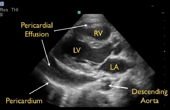
  1. Seif D, Perera P, Mailhot T, Riley D, Mandavia D. Bedside ultrasound in resuscitation and the rapid ultrasound in shock protocol. Crit Care Res Pract. 2012;2012:503254. (License: CC BY-3.0)
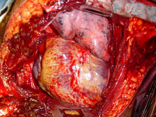
  2. Cothren CC, Moore EE. Emergency department thoracotomy for the critically injured patient: Objectives, indications, and outcomes. World J Emerg Surg. 2006 Mar 24;1:4. (License: CC BY-2.0)